Introduction
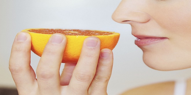
Smell and taste are generally classified as visceral senses because of their close association with gastrointestinal function. Physiologically, they are related to each other. The flavors of various foods are in large part a combination of their taste and smell. Consequently, food may taste “different” if one has a cold that depresses the sense of smell. Both smell and taste receptors are chemoreceptors that are stimulated by molecules in solution in mucus in the nose and saliva in the mouth. Because stimuli arrive from an external source, they are also classified as exteroceptors.
Smell
The olfactory sensory neurons are located in a specialized portion of the nasal mucosa, the yellowish pigmented olfactory epithelium. In dogs and other animals in which the sense of smell is highly developed (macrosmatic animals), the area covered by this membrane is large; in microclimatic animals, such as humans, it is small. In humans, it covers an area of 5 cm2 in the roof of the nasal cavity near the septum. The human olfactory epithelium contains 10 to 20 million bipolar olfactory sensory neurons interspersed with glia-like supporting (sustentacular) cells and basal stem cells.
Read More: thenewspointof.net
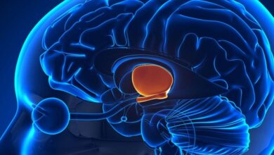
Olfactory bulbs
In the olfactory bulbs, the axons of the olfactory sensory neurons (first cranial nerve) contact the primary dendrites of the mitral cells and tufted cells to form anatomically discrete synaptic units called olfactory glomeruli.
Olfactory cortex
The axons of the mitral and tufted cells pass posteriorly through the lateral olfactory stria to terminate on apical dendrites of pyramidal cells in five regions of the olfactory cortex: anterior olfactory nucleus, olfactory tubercle, piriform cortex, amygdala, and entorhinal cortex. From these regions, information travels directly to the frontal cortex or via the thalamus to the orbitofrontal cortex. Conscious discrimination of odors is dependent on the pathway to the orbitofrontal cortex. The orbitofrontal activation is generally greater on the right side than the left; thus, cortical representation of olfaction is asymmetric
Olfactory thresholds & discrimination
The olfactory epithelium is covered by a thin layer of mucus secreted by the supporting cells and Bowman glands, which lie beneath the epithelium. The mucus bathes the odorant receptors on the cilia and provides the appropriate molecular and ionic environment for odor detection.
Taste
The specialized sense organ for taste (gustation) consists of approximately 10,000 taste buds, which are ovoid bodies measuring 50–70 μm. There are four morphologically distinct types of cells within each taste bud: basal cells, dark cells, light cells, and intermediate cells. The latter three cell types are all referred to as Type I, II, and III taste cells. They are the sensory neurons that respond to taste stimuli or tastants. The three cell types may represent various stages of differentiation of developing taste cells, with the light cells being the most mature. Alternatively, each cell type might represent different cell lineages.
Summary
Olfactory sensory neurons, supporting (sustentacular) cells, and basal stem cells are located in the olfactory epithelium within the upper portion of the nasal cavity.The cilia located on the dendritic knob of the olfactory sensory neuron contain odorant receptors which are coupled to heterotrimeric G proteins.