Introduction
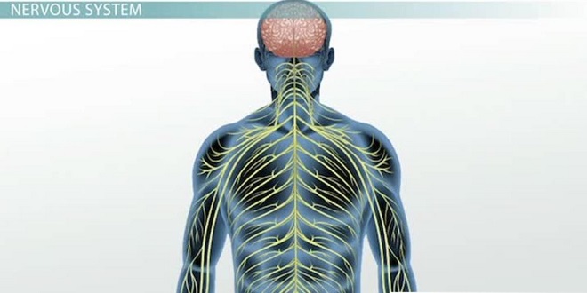
The autonomic nervous system (ANS) is the part of the nervous system that is responsible for homeostasis. Except for skeletal muscle, which gets its innervation from the somatomotor nervous system, innervation to all other organs is supplied by the ANS. Nerve terminals are located in smooth muscle (eg, blood vessels, gut wall, urinary bladder), cardiac muscle, and glands (eg, sweat glands, salivary glands). Although survival is possible without an ANS, the ability to adapt to environmental stressors and other challenges is severely compromised.
Anatomic organization of autonomic outflow
The cell bodies of the preganglionic neurons are located in the inter mediolateral column (IML) of the spinal cord and in the motor nuclei of some cranial nerves. In contrast to the large diameter and rapidly conducting α-motor neurons, preganglionic axons are small-diameter, militated, relatively slowly conducting B fibers. A preganglionic axon diverges to an average of eight or nine postganglionic neurons. In this way, autonomic output is diffused. The axons of the postganglionic neurons are mostly unmyelinated C fibers and terminate on the visceral effectors.
Sympathetic division
In contrast to α-motor neurons, which are located at all spinal segments, sympathetic preganglionic neurons are located in the IML of only the first thoracic to the third or fourth lumbar segments. This is why the sympathetic nervous system is sometimes called the thoracolumbar division of the ANS. The axons of the sympathetic preganglionic neurons leave the spinal cord at the level at which their cell bodies are located and exit via the ventral root along with axons of α- and γ-motor neurons.
Parasympathetic division
The parasympathetic nervous system is sometimes called the craniofacial division of the ANS because of the location of its preganglionic neurons. The parasympathetic nerves supply the visceral structures in the head via the coulometer, facial, and glossopharyngeal nerves, and those in the thorax and upper abdomen via the vague nerves. The sacral outflow supplies the pelvic viscera via branches of the second to fourth sacral spinal nerves. Parasympathetic preganglionic fibers synapse on ganglia cells clustered within the walls of visceral organs; thus these parasympathetic postganglionic fibers are very short.
Chemical transmission at autonomic junctions
The first evidence for chemical neurotransmission was provided by a simple yet dramatic study of heart rate control by the parasympathetic nervous system performed by Otto Loewi in 1920 (Clinical Box 17–2).
Responses of effector organs to autonomic nerve impulses
The effects of stimulation of the noradrenergic and cholinergic postganglionic nerve fibers are indicated. These findings point out another difference between the ANS and the somatomotor nervous system. The release of acetylcholine by α-motor neurons only leads to the contraction of skeletal muscles. In contrast, the release of acetylcholine onto smooth muscles of some organs leads to contraction (eg, walls of the gastrointestinal tract) while release onto other organs leads to relaxation (eg, sphincters in the gastrointestinal tract). The only way to relax a skeletal muscle is to inhibit the discharges of the α-motor neurons; but for some targets innervated by the ANS, one can shift from contraction to relaxation by switching from activation of the parasympathetic nervous system to activation of the sympathetic nervous system.
Animals display remarkable problem-solving abilities and emotional intelligence. Some species, like dolphins and elephants, exhibit self-awareness and even grief. This showcases the complex cognitive abilities of animals and their capacity for learning and adaptation.