Introduction
Receptors for two sensory modalities, hearing and equilibrium, are housed in the ear. The external ear, the middle ear, and the cochlea of the inner ear are concerned with hearing. The semicircular canals, the utricle, and the saccule of the inner ear are concerned with equilibrium. Receptors in the semicircular canals detect rotational acceleration, receptors in the utricle detect linear acceleration in the horizontal direction, and receptors in the saccule detect linear acceleration in the vertical direction. The receptors for hearing and equilibrium are hair cells, six groups of which are present in each inner ear: one in each of the three semicircular canals, one in the utricle, one in the saccule, and one in the cochlea.
Anatomic considerations
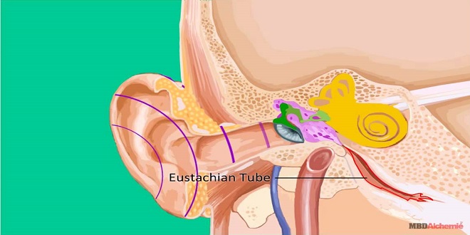
The external ear funnels sound waves to the external auditory meatus. In some animals, the ears can be moved like radar antennas to seek out sound. From the external auditory meatus, sound waves pass inward to the tympanic membrane (eardrum)
Read More: themakernewsz.com
Inner ear
The inner ear (labyrinth) is made up of two parts, one within the other. The bony labyrinth is a series of channels in the petrous portion of the temporal bone. Inside these channels, surrounded by a fluid called perilymph, is the membranous labyrinth. This membranous structure more or less duplicates the shape of the bony channels. It is filled with a K+-rich fluid called endolymph, and there is no communication between the spaces filled with endolymph and those filled with perilymph.
Utricle & saccule
Within each membranous labyrinth, on the floor of the utricle, is an otolithic organ (macula). Another macula is located on the wall of the saccule in a semivertical position. The maculae contain supporting cells and hair cells, surmounted by an otolithic membrane in which are embedded crystals of calcium carbonate, the otoliths. The otoliths, which are also cconia or ear dust, range from 3 to 19 μm in length in humans and are denser than the end lymph. The processes of the hair cells are embedded in the membrane. The nerve fibers from the hair cells join those from the cristae in the vestibular division of the eighth cranial nerve.
Hair cells
As noted above, the sensory receptors in the ear consist of six patches of hair cells in the membranous labyrinth. The hair cells in the organ of Corte signal hearing; the hair cells in the utricle signal horizontal acceleration; the hair cells in the saccule signal vertical acceleration; and a patch in each of the three semicircular canals signal rotational acceleration. These hair cells have a common structure. Each is embedded in an epithelium made up of supporting cells, with the basal end in close contact with afferent neurons. Projecting from the apical end are 30 to 150 rod-shaped processes, or hairs.
Summary
The external ear funnels sound waves to the external auditory meatus and tympanic membrane. From there, sound waves pass through three auditory icicles (malleus, incus, and stapes) in the middle ear. The inner ear, or labyrinth, contains the cochlea and organ of Corte.
The hair cells in the organ of Corti signal hearing. The stereocilia provide a mechanism for generating changes in membrane potential proportional to the direction and distance the hair moves. Sound is the sensation produced when longitudinal vibrations of air molecules strike the tympanic membrane.
Understanding the conjugation of verbs is essential in mastering a language. It involves various verb forms and tenses, which convey the timing and completion of actions. Additionally, mood and voice variations add further complexity to verb conjugation. The nuances of shabd roop encompass the subtleties of language, requiring attention to detail and practice for fluency.